The retina is a transparent tissue located inside the eye. The retina is similar to the film in the back of a camera. It receives visual images and transmits the information to the brain via the optic nerve.
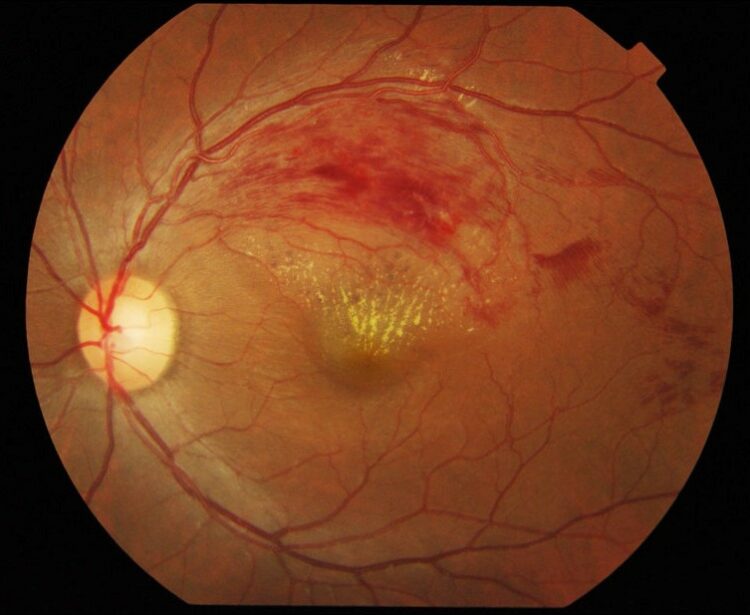
The retina receives its nourishment from tiny arteries. The blood in these arteries passes through small capillaries and eventually passes back to the heart via the retinal veins. In some individuals, one of the retinal veins may become plugged and this will cause sludging within the capillaries “upstream” from the obstruction. This branch retinal vein occlusion is, effectively, a localized “mini stroke” of the eye.
There may be mild to moderate loss of vision following a branch retinal vein occlusion. The loss of vision is due either to swelling of the retina or bleeding within the eye. In some patients, the vision may improve spontaneously. In cases of retinal swelling, if the vision remains poor for a period of weeks or months, your doctor may suggest an injection of intraocular Kenalog or, less often, intraocular Avastin. In addition, a laser treatment may be suggested following these injections. The purpose of this treatment is to prevent progressive visual loss and to attempt to provide improvement in vision. Often, repeat injections are required. In cases of bleeding within the eye, extensive laser treatment may help prevent recurrent hemorrhages. Rarely, surgery is advised to remove the blood.
The most common cause of branch retinal vein occlusion is hardening of the arteries (atherosclerosis) and high blood pressure. If high blood pressure is present, treatment of hypertension is indicated to prevent additional mini strokes from developing.