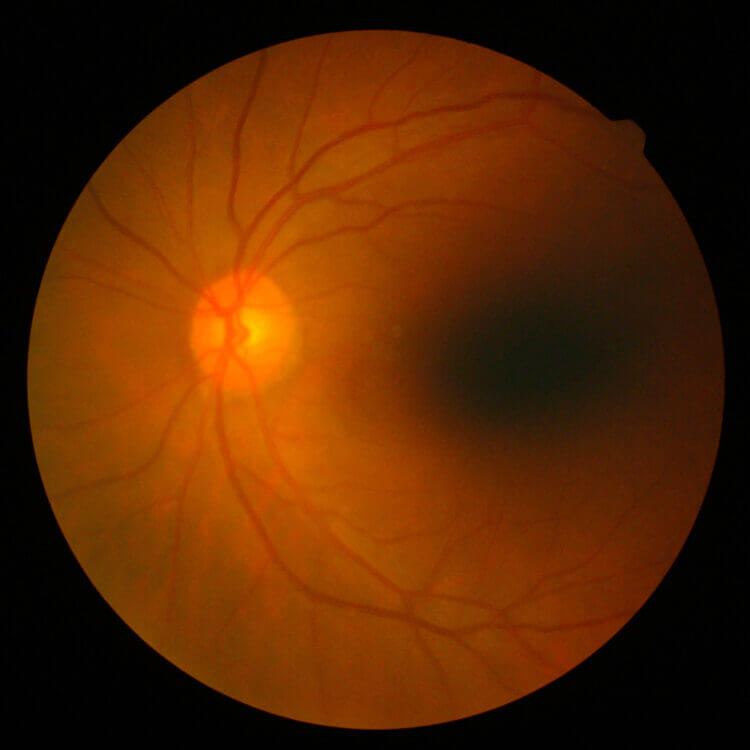
A retinal detachment occurs when the retina is lifted up and separates from the back surface of the eye. There are three types of retinal detachments. The most common type occurs when fluid seeps underneath the sensory layer of the retina, causing the layer of the retina to separate. This type of detachment occurs more frequently in people who are nearsighted, have had eye surgery or have sustained a serious injury to the eye.
The second most common type of detachment appears when the vitreous gel and scar tissue contract and pull on the retina causing it to lift up. This type of detachment, known as a traction retinal detachment, is more likely to occur in a patient with diabetes.
The third type arises when fluid collects under the retina causing it to separate from the back surface of the eye. This usually occurs from swelling and bleeding associated with another disease affecting the eye.
Symptoms
Symptoms of a retinal detachment may include a dark shadow in the field of vision, a veil or curtain appearance obstructing the vision, “wavy” or “watery” vision, light flashes, a shower of floaters resembling spots or spider webs and/or a sudden decrease in vision. If you are experiencing any of these conditions, contact your eye doctor immediately for an evaluation.
Treatment
In order for a patient to regain any vision, surgery must be performed to repair the retinal detachment. Possible treatments include Scleral Buckling surgery, Pneumatic Retinopexy or Vitrectomy surgery. The appropriate treatment will be determined by your doctor based on the type and severity of your condition.
